News, Events, & Announcements
April 17th, 2026
Check out our instagram page for fun images and little scientific tidbits:
https://www.instagram.com/osu_electron_microscopy/
April 14th, 2026
Rebecca just completed a very nice PowerPoint on Tissue prep for TEM. Please see this link. And remember, we are here to help with tissue prep - particularly cutting and staining - but this guide will give you a good idea of where to start.
April 13, 2026
Thanks for coming out to the open house. We showed off lots of nice samples and 3-D data sets. If you missed it, come by and see us and we can show you what we went through.
March 18 , 2026
We are having an open house. Friday, April 10th from 2-4 pm in LPSC room 145. Enjoy some finger food and refreshments while you talk with faciity staff and or your colleagues. Many 10 minute demos are planned on the Titan TEM Rebecca , the Helios (auto slicenview) Pete, and our 3-D reconstruction capability with our student helper Carmen.
March 16, 2026
The power outage Saturday took down our Quanta 600. It should be back by March 20th. We have not been super busy due to finals and have been accomodating everyone on the Helios and Q3D as both of those are up and running well.
Feb 4, 2026
We are in the process of crafting proposals to replace the now aging Q600 and Q3D in the next year! The Q3D with a plasma FIB and the Q600 with a more modern SEM with a fast EDS and a Navcam for ease of use.
If you have recently trained with us or have interacted with us, please fill out a survey: Survey link
We have not had an open house for a while. Look for one coming soon. We will have finger food and refreshments.
Sept 19, 2025
I just had a chance to add the wonderful, Titan training manual to our webpage. If you have not seen it, please check it out. It has full ray tracings of the optics systems and gives many helpful hints such as Gun Lens 8, spot 6 for fine, nanobeam diffraction. I just saved a register using those Gun Lens 6, and spot 6, CA 50 nano diffraction settings. Its never too late to learn something new.
Aug 1, 2025
Registration for our continuing ed. class, "Hands on Electron Microscopy" is now open. https://workspace.oregonstate.edu/course/hands-on-electron-microscopy-course
July 29, 2025
Welcome to the new fiscal year. We purchased a new automated compressor for the Quanta 600 and it works: no more having un expected vents during your Q600 sessions. The Q3D electron column is being rebuilt, new source, new power supply and new aperture controller. The sum of these repairs is over $60k . Come second week of August we expect it to be fully online again for your needs.
We have also implemented a survey. Tell us how we are doing.
April 22, 2025
Rebecca and Pete still continue to make very good to world class samples and consult and teach students. Pete has now passed the 1000 FIB samples and continues to receive a steady stream of sample making requests , some from out of state. Rebecca, although only here for two years makes wonderful samples that have visible mitochondria, membranes and other ultra structures. The Titan TEM just received a preventative maintenance. And the Gatan, Rio side mount camera continues to be very easy to use and provide 2x to 3x the contrast of the lower Ultra Scan camera. The Ultra Scan camera with its 3 times longer camera length is still most suitable for high magnifications. Note, the Titan is not image or probe corrected.
The continuing education course is back by popular demand and look for it in the Fall.
July 31, 2024
Our continuing education course, "Hands on Electron Microscopy," is now open for registration. It is a 10 week course one lecture per week. And features 30 minutes of hands on demos every session.
https://workspace.oregonstate.edu/course/hands-on-electron-microscopy-c…
July 12th, 2024
We have just confirmed that Gatan will be installing a new side mount, "Rio" camera in the OSU Titan the week of August 5th. This is a CMOS camera with fast frame rates: 20 FPS at full resolution and 200 FPS at 1k by 1K resolution. It also has 4D STEM capability, look for the 4D STEM pack at a later date. Our strategy is to update our Titan TEM with the newest of technology as it becomes available.
Feb 24th , 2024
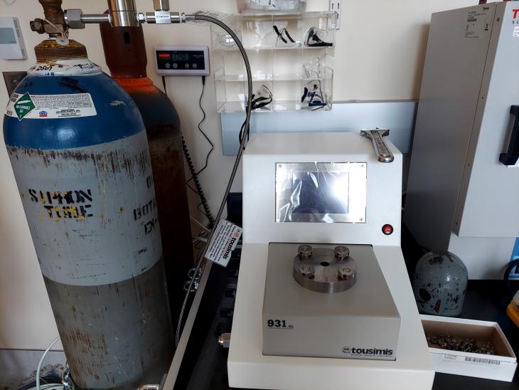
The EM Facility has recently acquired a new Critical Point Dryer for sample preparation!
January 25th 2024
Happy New Year from the EM Facility! We hope that you’ve all had a great first month of the new year!
For today’s announcement, we highly recommend watching a wonderful live demonstration of our FIB FIG system from our facility director, Dr. Peter Eschbach. Check out this neat technique via the link below!
https://thermofisher.app.box.com/s/4b5gu74q81xyze37zmhl4r4yxbsn55zj/file/1425318591783
Closed January 15th (Martin Luther King Jr. Day)
The EM Facility will be closed on January 15th for Martin Luther King Jr. Day.
December 22
Announcement's:
- The EM facility be closed between 25-29th.
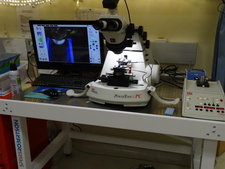
- We've acquired a lovely new table, housing our Ultra Microtome as shown below.
We hope that you all have a wonderful winter break! Happy Holidays!!
RELMS , our scheduling system is in full swing and it has really helped streamline our billing. As of April 1st, RELMS was the platform that will be used for booking Quanta 600, Quanta 3D, Titan and Helios.
At this time masks are not required
And as always, we strive to treat all nationalities and ethnic backgrounds with respect and equity.